MEDED
ASSESSMENTS
Create. Analyze. Share.
Error message
success message
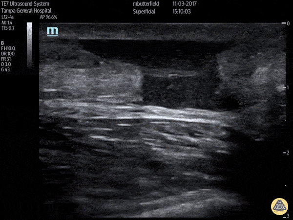
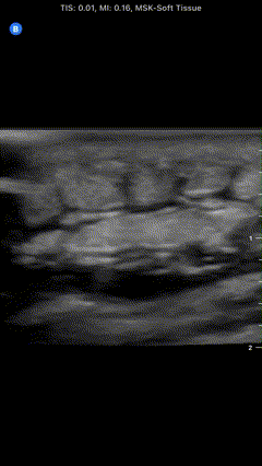
Which is seen in the following clip of an Achilles tendon?
1
1 pts
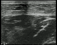
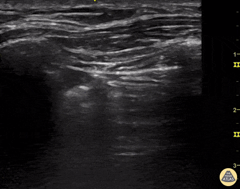
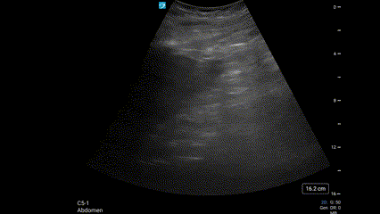
Which is seen in this clip of subcutaneous tissue?
2
1 pts
Which of the following represents a common artifact in musculoskeletal scanning, when the probe is not perpendicular to the structure and causes hypoechoic areas due to sound waves not reflecting back?

3
1 pts
Below are long- and short-axis views of a patient's inferior vena cava. In the long-axis view, the patient performed a sniff maneuver. Which of the following correctly interprets these images?

4
1 pts
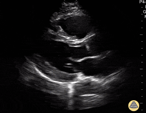
A patient presents with acute right knee pain. The suprapatellar ultrasound image below is obtained. Which is seen?

5
1 pts
Which of the following probes should be used for cardiac examination?
6
1 pts
Which of the following is true regarding ultrasound probe selection?
7
1 pts
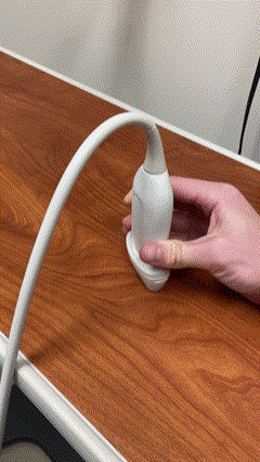
Which of the following terms correctly describes moving the ultrasound probe along the long axis either towards or away from the probe indicator as shown?
8
1 pts
Which of the following best explains the findings seen above the diaphragm?

9
1 pts
Which is seen in the image below?
10
1 pts
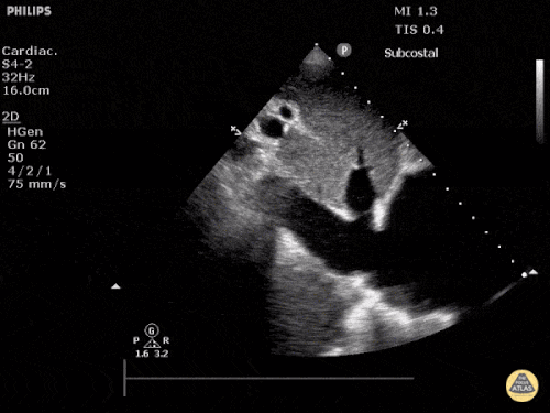
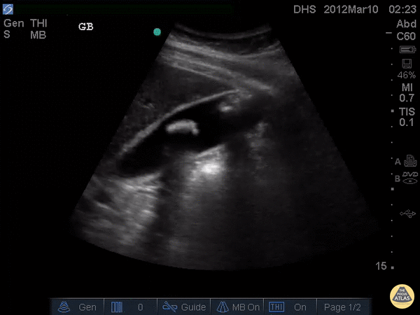
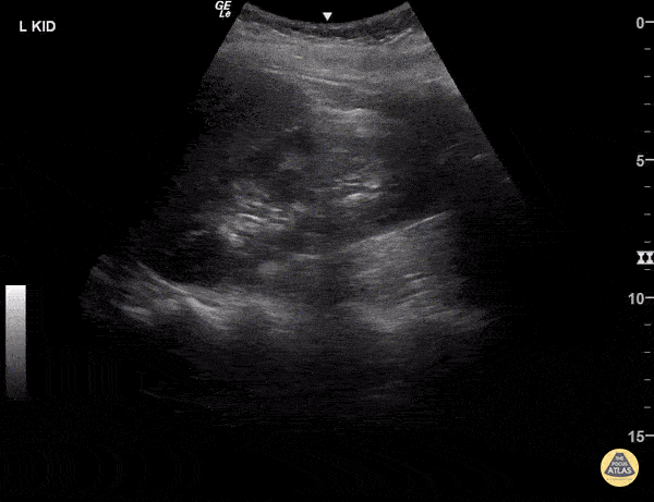
What is present?
11
1 pts
Which finding is suggested by the image below?
12
1 pts
Which is suggested by the below image, in which the IVC measures 2.4cm?
13
1 pts
Which are suggested by the images below, obtained from the same patient?
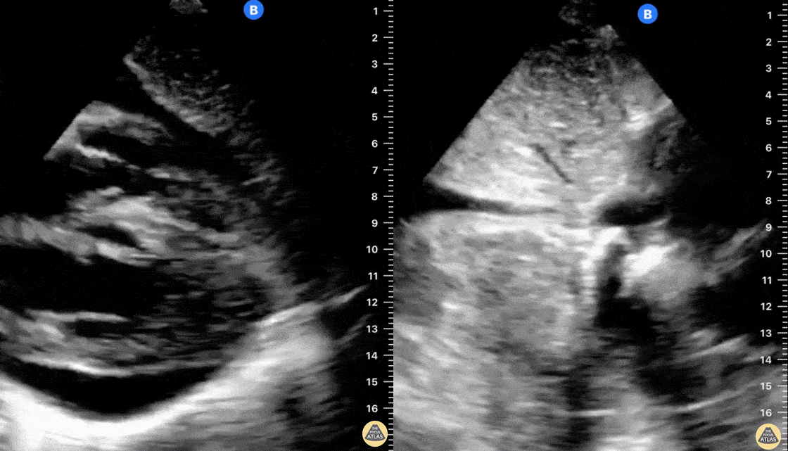
14
1 pts
Which is present in the image below?
15
1 pts
Which is suggested by the image below?
16
1 pts
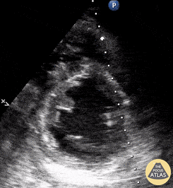
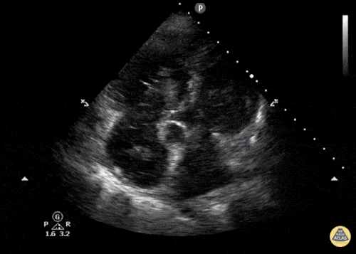
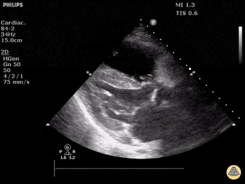
Which valve appears abnormal?
17
1 pts
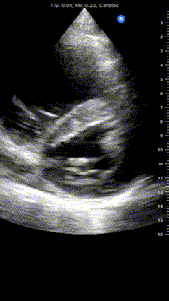
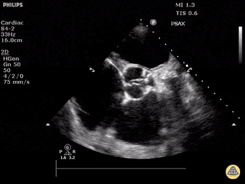
Which of the following is demonstrated?

18
1 pts
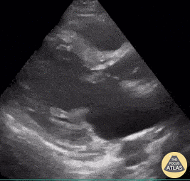
Which of the following is seen in the image below?
19
1 pts
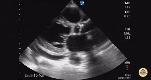
Which is suggested by the image below?
20
1 pts
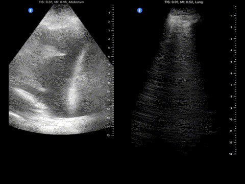
The following images are obtained from the same patient's right lung (image on the left) and left lung (image on the right). Which of the following is favored based on this pulmonary POCUS?
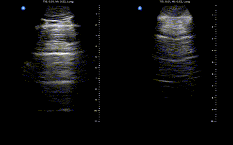
21
1 pts
Which of the following is most likely based on the below pulmonary POCUS findings?
22
1 pts
A trainee sends you this image that they obtained from the posterolateral aspect of a patient's chest. They were assessing for pulmonary or pleural abnormalities. Which of the following best describes the findings?
23
1 pts
How many B lines are required to be abnormal?
24
1 pts
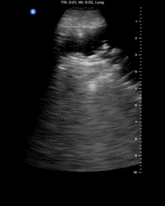
Which of the following best describes this imaging finding?
25
1 pts
The images below are thoracic POCUS images from both sides of the same patient (right lung on the left, left lung on the right). These pulmonary POCUS findings would most likely be associated with which diagnosis?
26
1 pts
Which of the following etiologies of shock is most suggested by hyperdynamic left ventricular function, a reduced TAPSE, and a predominant A-line pattern?
27
1 pts
This video is most suggestive of which shock type?
28
1 pts
Which of the following is most specific for cardiac tamponade?
29
1 pts
This ultrasound video is most suggestive of which type of shock?
30
1 pts
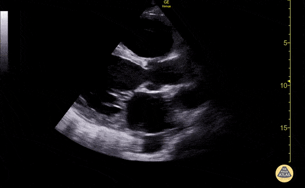
An IVC diameter of 2.2cm and >50% collapsibility is most consistent with a central venous pressure of...
31
1 pts
Which is the most appropriate single method to assess for proximal DVT?
32
1 pts
The vessel indicated by the red dot is the...

33
1 pts
Which of the following best describes these skin ultrasound findings?
34
1 pts
Which of the following are most consistent with the skin and soft tissue ultrasound findings below?

35
1 pts
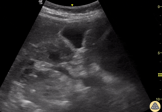
Which of the following can be directly visualized by a standard FAST exam?
36
1 pts
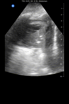
Based solely on the available image, which of the following is the best interpretation of this FAST exam?
37
1 pts
The below image is most consistent with...
38
1 pts
Which of the following structures is indicated by the red arrow?
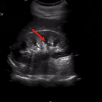
39
1 pts
Which best describes the image below?
40
1 pts
Which of the following best describes the findings shown?
41
1 pts
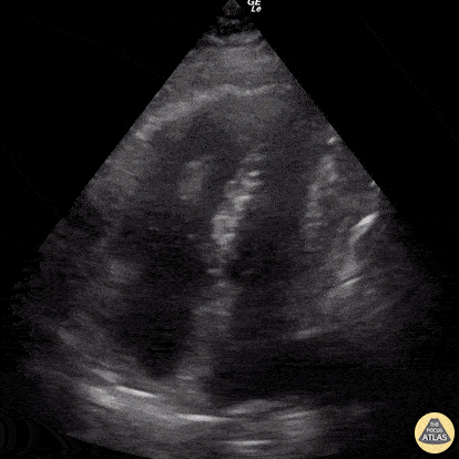
Which of the following is true regarding E-point septal separation (EPSS)?
42
1 pts